Other examples include the relationship between the anterior end of the other 11 ribs and the costal cartilage. Examples include the thoracic cage, such as the first sternocostal joint: the first rib is joined to the manubrium by its costal cartilage.


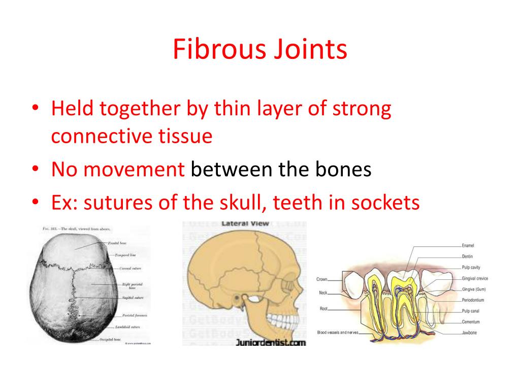
Permanent synchondroses function to connect bones without movement as a synarthrosis joint. Other temporary synchondroses join the ilium, ischium, and pubic bones of the hip over time, these also fuse into a single hip bone.Ī permanent synchondrosis does not ossify with age it retains its hyaline cartilage. Eventually, when all the hyaline cartilage has ossified, the bone is done lengthening ad the diaphysis and epiphysis fuse in synostosis. Over time, the cartilaginous plate expands and is replaced by bone, adding to the diaphysis. The epiphyseal plate connects the diaphysis (shaft of the bone) with the epiphysis (end of the bone) in children. Ī synchondrosis, or primary cartilaginous joint, only involves hyaline cartilage and can be temporary or permanent.Ī temporary synchondrosis is an epiphyseal plate (growth plate), and it functions to permit bone lengthening during development. The interosseous membranes of the leg and forearm are also areas of muscle attachment. For example, the tibiofibular syndesmosis primarily provides strength and stability to the leg and ankle during weight-bearing however, the antebrachial interosseous membrane of the radioulnar syndesmosis permits rotation of the radius bone during forearm movements. All syndesmoses are amphiarthroses, but each specific syndesmosis joint permits a different degree of movement. Of the fibrous joints, sutures and gomphoses are found only in the skull and the teeth, respectively.Ī syndesmosis, an amphiarthrosis joint, and the third type of fibrous joint maintain integrity between long bones and resists forces that attempt to separate the two bones. Within these categories, each specific joint type (suture, gomphosis, syndesmosis, synchondrosis, symphysis, hinge, saddle, planar, pivot, condyloid, ball, and socket) has a specific function in the body. The histological and functional classification schemes offer a broad understanding of joints.

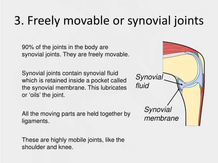
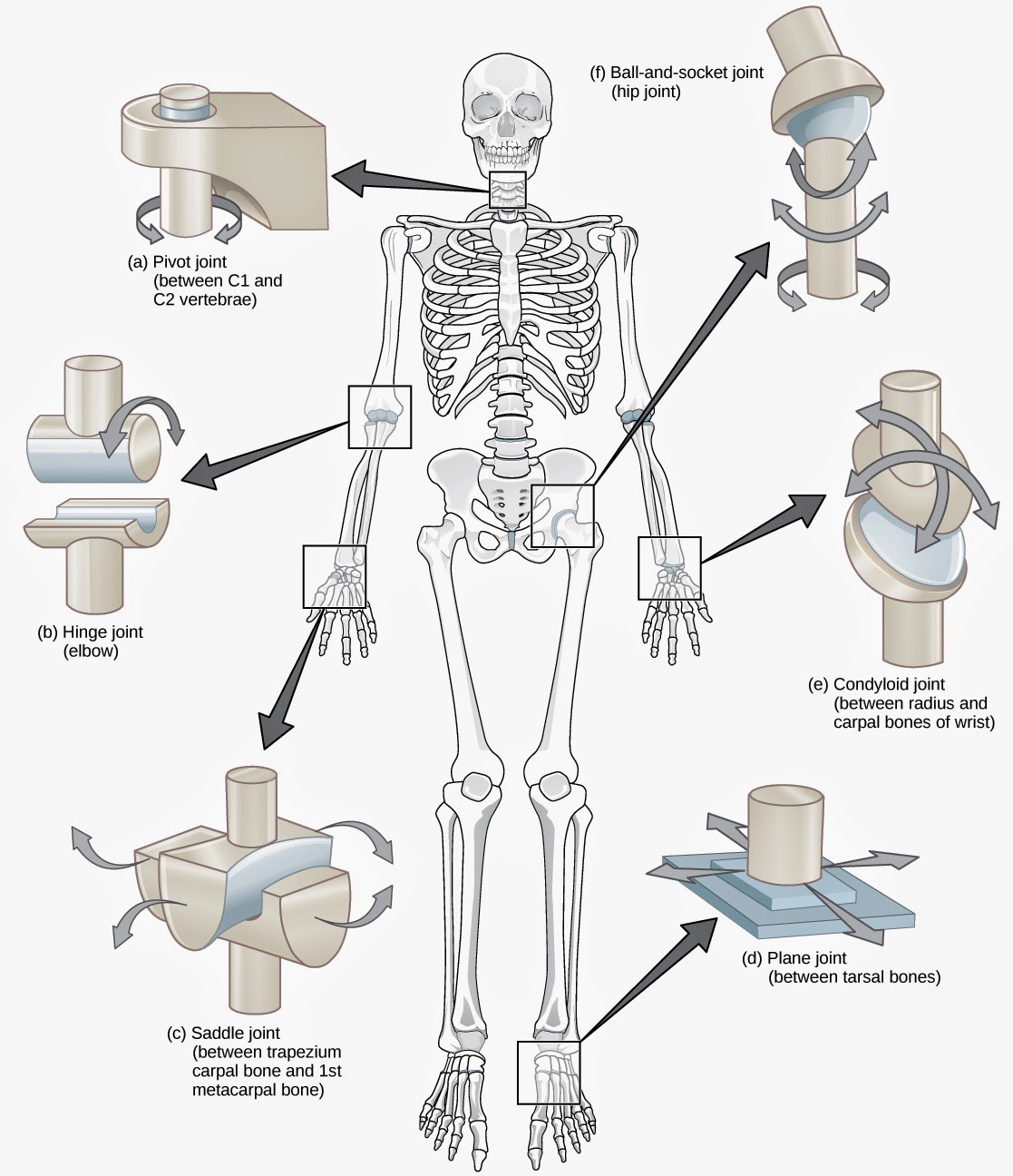
There are six such classifications: hinge (elbow), saddle (carpometacarpal joint), planar (acromioclavicular joint), pivot (atlantoaxial joint), condyloid (metacarpophalangeal joint), and ball and socket (hip joint). Synovial joints are often further classified by the type of movements they permit. Some synovial joints also have associated fibrocartilage, such as menisci, between articulating bones. The articular cartilage and the synovial membrane are continuous. Hyaline cartilage forms the articular cartilage, covering the entire articulating surface of each bone. The joint cavity contains synovial fluid, secreted by the synovial membrane (synovium), which lines the articular capsule. The cavity is surrounded by the articular capsule, which is a fibrous connective tissue that is attached to each participating bone just beyond its articulating surface.

Its joint cavity characterizes the synovial joint. Synovial joints are freely mobile (diarthroses) and are considered the main functional joints of the body. These joints are slightly mobile (amphiarthroses). The secondary cartilaginous joint, also known as symphysis, may involve either hyaline or fibrocartilage.


 0 kommentar(er)
